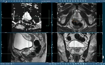

Precision Imaging: Radiology Assessments Unveiled
Advancements in radiology have revolutionized medical diagnostics, making radiology assessments an indispensable tool for precision imaging. From detecting subtle abnormalities to guiding complex medical procedures, radiology assessments play a crucial role in unraveling the intricacies of the human body.
The Essence of Radiology Assessments
Radiology assessments involve the use of medical imaging techniques to visualize the internal structures of the body. This encompasses various modalities, including X-rays, computed tomography (CT), magnetic resonance imaging (MRI), ultrasound, and nuclear medicine. Each modality serves unique purposes, allowing healthcare professionals to obtain detailed insights into different anatomical regions.
Unveiling Anomalies with X-rays
X-rays remain a fundamental component of radiology assessments. They are particularly effective in visualizing bones and detecting fractures, joint abnormalities, or certain lung conditions. The speed and simplicity of X-ray imaging make it a valuable tool for quick assessments in emergency situations or routine examinations.
Cross-Sectional Precision with CT Scans
Computed tomography (CT) scans provide cross-sectional images of the body with exceptional precision. CT assessments are instrumental in identifying and characterizing soft tissue abnormalities, vascular conditions, and abnormalities within the chest and abdomen. The detailed images obtained through CT scans aid in accurate diagnosis and treatment planning.
Magnetic Resonance Imaging (MRI) for Detailed Visualization
MRI is renowned for its ability to provide detailed images of soft tissues, organs, and the nervous system. Radiology assessments using MRI are crucial for evaluating conditions such as brain disorders, spinal cord abnormalities, and musculoskeletal injuries. The non-invasive nature of MRI makes it a preferred choice for many diagnostic scenarios.
Dynamic Insights with Ultrasound Imaging
Ultrasound imaging offers dynamic insights into the body’s structures and functions. It is commonly used for assessing pregnancy, cardiovascular conditions, abdominal organs, and the musculoskeletal system. The real-time nature of ultrasound enables healthcare professionals to observe organ function and blood flow dynamically.
Nuclear Medicine: Unraveling Cellular Activity
Nuclear medicine involves the use of radioactive tracers to visualize cellular activity within the body. This specialized form of radiology assessment is valuable for studying organ function, detecting tumors, and assessing conditions such as thyroid disorders. Nuclear medicine provides unique insights into the molecular and metabolic processes occurring within tissues.
Guiding Interventions with Fluoroscopy
Fluoroscopy is a real-time imaging technique that plays a pivotal role in guiding medical procedures. Whether it’s visualizing the movement of contrast agents during gastrointestinal studies or guiding minimally invasive interventions, fluoroscopy provides dynamic, live imaging that enhances the precision of medical procedures.
3D Reconstruction for Surgical Planning
Advanced radiology assessments include techniques for three-dimensional (3D) reconstruction of medical images. This is particularly valuable in surgical planning, where detailed 3D models of organs or structures aid surgeons in understanding complex anatomy and planning precise interventions.
Artificial Intelligence Integration for Enhanced Analysis
The integration of artificial intelligence (AI) in radiology assessments has further elevated diagnostic capabilities. AI algorithms can assist in image analysis, pattern recognition, and even help predict disease outcomes. This integration enhances the efficiency and accuracy of radiological interpretations, ultimately benefiting patient care.
Ensuring Patient Safety and Minimizing Radiation Exposure
As radiology assessments play a central role in diagnostics, ensuring patient safety is paramount. Modern radiological technologies prioritize minimizing radiation exposure while maintaining diagnostic efficacy. Strict safety protocols, dose optimization techniques, and advancements in imaging technology contribute to the overall safety of radiology assessments.
Conclusion: Precision and Progress in Radiology Assessments
In conclusion, radiology assessments have undergone remarkable advancements, providing healthcare professionals with unparalleled precision in diagnostics. From traditional X-rays to cutting-edge technologies like AI integration, radiology continues to evolve, ensuring accurate and comprehensive insights into the human body. The synergy of technology, expertise, and a commitment to patient well-being defines the landscape of modern radiology assessments.
For more information on Radiology Assessments, visit cloudfeed.net.
